
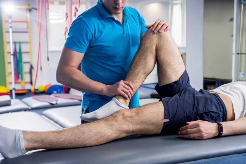

Excess body weight places continuous mechanical stress on the musculoskeletal system, particularly the weight-bearing joints of the lower extremities and spine. While lifestyle modifications frequently focus on cardiovascular markers or metabolic improvements, joint preservation remains one of the most immediate structural benefits of weight control. Combining strategic body mass reduction with guided physical therapy establishes a biomechanical environment where cartilage degradation slows, systemic inflammation drops, and functional mobility returns.
The Biomechanics of Weight and Joint Load
The human skeleton operates on a system of levers and fulcrums. Because of these leverage dynamics, the force exerted across joints during movement is significantly higher than an individual’s static scale weight.
-
The Knee Joint: During normal flat-ground walking, the force transmitted through the knee equals roughly three to four times total body mass. Climbing stairs or running increases this impact to six to eight times body mass. Consequently, shedding ten pounds unloads thirty to forty pounds of kinetic force from the patellofemoral and tibiofemoral joints with every single step.
-
The Hip Joint: The hip functions as a ball-and-socket joint stabilized by the gluteal complex and pelvic ligaments. Single-leg stance during gait amplifies pressure on the acetabulum to nearly three times body weight, making hip cartilage highly vulnerable to accelerated wear under surplus mass.
-
The Lumbar Spine: Extra weight, especially when carried anteriorly around the abdomen, shifts the center of gravity forward. This anterior displacement increases shear stress on the lumbar intervertebral discs and forces the posterior paraspinal muscles into chronic hyper-contraction to maintain an upright posture.
-
The Ankle and Foot: The subtalar joint and plantar fascia absorb primary ground reaction forces. Surplus mass flattens the medial longitudinal arch, triggering overpronation, altered tibial internal rotation, and cascading torque upward through the kinetic chain.
Mechanical Wear Versus Metabolic Inflammation
For decades, degenerative joint conditions such as osteoarthritis were classified exclusively as “wear and tear” disorders caused by pure friction and compressive loading. Modern clinical research shows that excess body mass harms joints through two distinct, concurrent mechanisms: direct mechanical overload and systemic biochemical inflammation.
Adipose tissue is an active endocrine organ, not an inert storage layer. Visceral and subcutaneous fat cells release bioactive molecules known as adipokines, which include leptin, resistin, and visfatin, along with pro-inflammatory cytokines like tumor necrosis factor-alpha and interleukin-6. These biochemical agents enter the bloodstream and trigger catabolic signaling inside synovial joint capsules. They degrade chondrocytes, dissolve the extracellular matrix of cartilage, and irritate the synovial lining.
This systemic biochemical attack explains why individuals carrying surplus weight experience elevated rates of osteoarthritis in non-weight-bearing regions, such as the distal interphalangeal joints of the hands. Reducing total adipose volume halts this inflammatory cascade, mitigating joint pain through biological pathways long before significant structural alterations appear on diagnostic imaging.
Why Weight Loss Alone Is Insufficient
Caloric reduction decreases downward mechanical pressure and dampens circulation-borne inflammatory cytokines, but it does not automatically repair years of compensatory movement patterns. Longstanding joint pain causes patients to adapt their gait, shorten their stride, shift weight away from painful limbs, and underutilize critical stabilizer muscles.
Sudden weight loss without structured physical rehabilitation often leads to:
-
Sarcopenia and Muscle Atrophy: Restrictive diets prompt the body to shed lean muscle tissue alongside adipose mass. Weaker quadriceps, hamstrings, and gluteal muscles leave the underlying joint surfaces exposed to unstable, unbuffered forces.
-
Persistent Biomechanical Faults: Removing weight does not teach the central nervous system how to activate dormant stabilizers. A patient may still walk with an antalgic limp or excessive pelvic tilt simply out of motor habit.
-
Tendon Laxity and Joint Instability: Rapid tissue loss can alter soft-tissue tension surrounding the joint capsule, yielding micro-instability if dynamic muscular control is absent.
The Role of Physical Therapy in Joint Rehabilitation
Physical therapy bridges the gap between passive weight reduction and active functional resilience. A physical therapist conducts comprehensive kinetic assessments to locate deficits in strength, joint range of motion, and balance, designing a customized regimen that protects healing articular cartilage.
Restoring Muscular Balance and Shock Absorption
Muscles act as dynamic shock absorbers. When the foot strikes the pavement, strong eccentric contraction of the quadriceps and calf complex dissipates impact energy before it reaches the subchondral bone. Physical therapists target specific stabilizing groups to optimize alignment:
-
Closed-Kinetic Chain Quadriceps Strengthening: Exercises such as shallow wall sits and leg presses build vastus medialis strength without provoking high shearing stresses under the patella.
-
Gluteal Complex Activation: Strengthening the gluteus medius prevents contralateral pelvic drop during gait, directly reducing valgus collapse at the knee.
-
Core and Pelvic Stabilization: Activating the transversus abdominis and multifidus stabilizes the lumbar spine, counteracting anterior pelvic tilt and unloading facet joints.
Low-Impact Conditioning Modalities
Cardiovascular exercise accelerates caloric expenditure, but high-impact routines exacerbate inflamed joints. Physical therapy introduces safe, non-concussive aerobic training:
-
Aquatic Therapy: Water provides natural buoyancy, offloading up to ninety percent of body weight depending on immersion depth, allowing active joint excursion without compressive penalty.
-
Recumbent Steppers and Stationary Cycling: Controlled, non-ballistic circular movements circulate synovial fluid across articular surfaces, delivering nutrients to cartilage without high peak impact loads.
-
Blood Flow Restriction Training: Utilizing pneumatic cuffs on target limbs allows patients to achieve muscular hypertrophy using loads as low as twenty percent of their one-rep maximum, avoiding joint strain altogether.
Creating a Sustainable Joint Health Program
Sustained joint health requires a progressive, three-stage continuum. The initial phase focuses on acute pain management, localized swelling control, and passive-to-active range of motion restoration. The intermediate phase shifts toward hypertrophy of primary stabilizing muscle groups and low-impact conditioning. The final phase emphasizes neuromuscular re-education, functional balance drills, and independent lifestyle execution.
Pairing steady caloric control with progressive resistance training yields compound physiological benefits. As fat mass drops, joint compression decreases; simultaneously, as lean muscle increases, structural support improves. This dual-action pathway halts cartilage breakdown, alleviates chronic discomfort, and restores long-term mobility.
Frequently Asked Questions
Does low-impact exercise burn enough calories to contribute directly to weight loss?
Yes. While high-impact routines expend slightly more calories per minute, low-impact alternatives such as swimming, rowing, and cycling can be sustained for longer durations with far less fatigue-related injury risk. Energy expenditure is primarily driven by duration and heart rate consistency, not joint impact.
How does systemic dehydration affect joint comfort during a fitness program?
Articular cartilage consists of approximately seventy to eighty percent water, supported by a matrix of proteoglycans. Insufficient hydration decreases the lubricating efficacy and compressive shock resistance of cartilage, elevating internal friction and worsening discomfort during movement.
Can physical therapy restore cartilage that has completely worn down?
Physical therapy cannot regrow articular cartilage that has reached bone-on-bone degradation. However, it can optimize the surrounding muscular support structure, expand joint space mechanically, and balance distribution loads to make the joint functional and pain-free without invasive interventions.
What is the difference between open-chain and closed-chain exercises for painful joints?
In open-chain exercises, the limb moves freely against resistance, such as a seated knee extension machine. In closed-chain exercises, the hand or foot stays planted against a fixed surface, such as a squat or step-down. Closed-chain exercises recruit multiple stabilizing muscle groups simultaneously and generate more natural, joint-protective forces.
Why does cold or damp weather frequently increase pain in compromised joints?
Drops in ambient barometric pressure allow the soft tissues and synovial fluid inside joint capsules to expand slightly. In an already inflamed or structurally tight joint, this micro-expansion places additional tension on localized sensory nerve endings, driving up pain perception.
Can rapid weight loss negatively impact bone mineral density around weight-bearing joints?
Yes. Rapid, highly restrictive weight loss, especially when unaccompanied by resistance training, can trigger a reduction in bone mineral density. The subchondral bone beneath articular cartilage requires mechanical signals from safe resistance training to preserve its density and internal architecture.
How do tight calf muscles affect the biomechanical alignment of the knee and hip?
Restricted flexibility in the gastrocnemius and soleus prevents normal forward movement of the shin bone during a stride. To bypass this limitation, the body compensates through premature heel-off or extreme foot overpronation, which introduces unwanted internal rotation through the knee and pelvic girdle.

Comments are closed.